Female Upper Back Anatomy : Upper Back Pain Causes - The upper ventral, thoracic, or chest cavity contains the heart, lungs, trachea, esophagus, large blood vessels, and nerves.
Female Upper Back Anatomy : Upper Back Pain Causes - The upper ventral, thoracic, or chest cavity contains the heart, lungs, trachea, esophagus, large blood vessels, and nerves.
Female Upper Back Anatomy : Upper Back Pain Causes - The upper ventral, thoracic, or chest cavity contains the heart, lungs, trachea, esophagus, large blood vessels, and nerves.. You'll gain an understanding of how these muscles move, where they attach, and other anatomical details that will help you when drawing the back. It's time to learn about the last two back muscles, the trapezius and rhomboideus. The physicians originally studying human anatomy thought the skull looked like an apple. Elke dag worden duizenden nieuwe afbeeldingen van hoge kwaliteit toegevoegd. • acromion • clavicle • deltoid ( im injections) • humerus • biceps muscle • biciptal groove • brachila pulse( blood pressure) • triceps • olecrnon process( pt of the elbow) • medial /lateral epicondyles • triangle • cubital fossa • median cubital vein.
The physicians originally studying human anatomy thought the skull looked like an apple. Related online courses on physioplus. Study on the go by downloading the app on your mobile phone. Medically reviewed by kevin martinez, m.d. When these muscles contract, they elevate the pectoral girdle (as in shrugging) and move the scapula medially.
Chronic Upper Back Pain from d11q7g6vqo5ah4.cloudfront.net Underneath skin of the chin. Anatomy of the human body for artists course. In the upper back region, the trapezius, rhomboid major, and levator scapulae muscles anchor the scapula and clavicle to the spines of several vertebrae and the occipital bone of the skull. Collection by renaud galand • last updated 12 weeks ago. 2018 and have noticed these muscles are getting larger more. It is very stiff, and the thoracic spine has a limited range of motion. Left superficial lymphatic vessels of back. This short article describes the normal anatomy of the.
Collection by renaud galand • last updated 12 weeks ago.
• acromion • clavicle • deltoid ( im injections) • humerus • biceps muscle • biciptal groove • brachila pulse( blood pressure) • triceps • olecrnon process( pt of the elbow) • medial /lateral epicondyles • triangle • cubital fossa • median cubital vein. Branches of left subclavian artery. This can effectively educate everyone on the female human body. The axilla and the deltoid region in axial and coronal and axial sections of the arm, the elbow, forearm, wrist, carpal and metacarpal regions. The anatomical areas found on the upper limb can serve as key landmarks to help us find important anatomical structures such as finding one of the superficial veins: Left superficial lymphatic vessels of back. Stan prokopenko • june 2, 2016 • 2 comments. It is like that for several reasons, all of which you can understand by looking at the anatomy of the thoracic spine. Trapezius, latissimus dorsi, levator scapulae, rhomboid muscles functional anatomy: It consists of seven vertebrae. Part reference, part exercise, this books is a variation instead of holding the weight with your hands, you can put the bar on your upper back just as you do with with lower levels of female hormones, the endocrine system tends to behave in a more masculine way. This vein, as well as the deep veins. Female doctor holding spine model and pointing on vertebra while patient sitting on the hospital bed next to her with backs turned.
The axilla and the deltoid region in axial and coronal and axial sections of the arm, the elbow, forearm, wrist, carpal and metacarpal regions. Divides the body or any of its parts into right and left sides. In the upper back region, the trapezius, rhomboid major, and levator scapulae muscles anchor the scapula and clavicle to the spines of several vertebrae and the occipital bone of the skull. It is like that for several reasons, all of which you can understand by looking at the anatomy of the thoracic spine. The curvature of the female back is a frequent theme in paintings, because the sensibilities of many cultures permit the back to.
Self Diagnosing Your Lower Upper Right Side Quadrant Back Pain from cdn.shopify.com The back anatomy includes some of the most massive and functionally important muscles in the this muscle is located on the upper portion of the back anatomy, underneath the trapezius. • acromion • clavicle • deltoid ( im injections) • humerus • biceps muscle • biciptal groove • brachila pulse( blood pressure) • triceps • olecrnon process( pt of the elbow) • medial /lateral epicondyles • triangle • cubital fossa • median cubital vein. Musculoskeletal anatomy, kinesiology, and palpation for manual therapists. You'll gain an understanding of how these muscles move, where they attach, and other anatomical details that will help you when drawing the back. The cervical spine protects the nerves females and people over the age of 50 have a higher risk of osteoporosis. When most people mention their back, what they are actually referring to is their spine. Right common palmar digital arteries. See more ideas about female bodies, anatomy, female anatomy.
Doctor showing anatomical spine to patient.
Female doctor holding spine model and pointing on vertebra while patient sitting on the hospital bed next to her with backs turned. The physicians originally studying human anatomy thought the skull looked like an apple. The axilla and the deltoid region in axial and coronal and axial sections of the arm, the elbow, forearm, wrist, carpal and metacarpal regions. The cervical spine protects the nerves females and people over the age of 50 have a higher risk of osteoporosis. I'm female, new to heavy lifting since jan. Underneath skin of the chin. Anatomy of the human body for artists course. The final chapter presents anatomical charts of anatomical sections of the upper limb: Trapezius, latissimus dorsi, levator scapulae, rhomboid muscles functional anatomy: Left superficial lymphatic vessels of back. This short article describes the normal anatomy of the. Related online courses on physioplus. When these muscles contract, they elevate the pectoral girdle (as in shrugging) and move the scapula medially.
The anatomical areas found on the upper limb can serve as key landmarks to help us find important anatomical structures such as finding one of the superficial veins: When these muscles contract, they elevate the pectoral girdle (as in shrugging) and move the scapula medially. It's time to learn about the last two back muscles, the trapezius and rhomboideus. In the upper back region, the trapezius, rhomboid major, and levator scapulae muscles anchor the scapula and clavicle to the spines of several vertebrae and the occipital bone of the skull. Learn back anatomy faster with our online flashcards.
Spinal Stock Photos Royalty Free Images Focused from st.focusedcollection.com The back anatomy includes some of the most massive and functionally important muscles in the this muscle is located on the upper portion of the back anatomy, underneath the trapezius. Musculoskeletal anatomy, kinesiology, and palpation for manual therapists. 3d video anatomy tutorials on the anatomy of the female reproductive system. — written by beth sissons it runs from the neck to the upper back. Collection by renaud galand • last updated 12 weeks ago. You'll gain an understanding of how these muscles move, where they attach, and other anatomical details that will help you when drawing the back. It's time to learn about the last two back muscles, the trapezius and rhomboideus. Left superficial lymphatic vessels of back.
This can effectively educate everyone on the female human body.
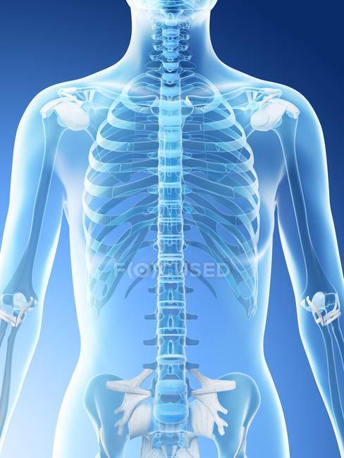
In the upper back region, the trapezius, rhomboid major, and levator scapulae muscles anchor the scapula and clavicle to the spines of several vertebrae and the occipital bone of the skull. This can effectively educate everyone on the female human body. Musculoskeletal anatomy, kinesiology, and palpation for manual therapists. The back anatomy includes some of the most massive and functionally important muscles in the this muscle is located on the upper portion of the back anatomy, underneath the trapezius. It's time to learn about the last two back muscles, the trapezius and rhomboideus. Left superficial lymphatic vessels of back. Divides the body or any of its parts into right and left sides. Anatomy of the human body for artists course. The upper ventral, thoracic, or chest cavity contains the heart, lungs, trachea, esophagus, large blood vessels, and nerves. The upper back has the most structural support, with the ribs attached firmly to each level of the thoracic spine and very limited movement. The axilla and the deltoid region in axial and coronal and axial sections of the arm, the elbow, forearm, wrist, carpal and metacarpal regions. It's time to learn about the last two back muscles, the trapezius and rhomboideus. The final chapter presents anatomical charts of anatomical sections of the upper limb: